Confidence and efficiency in structural heart interventions
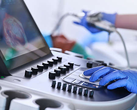
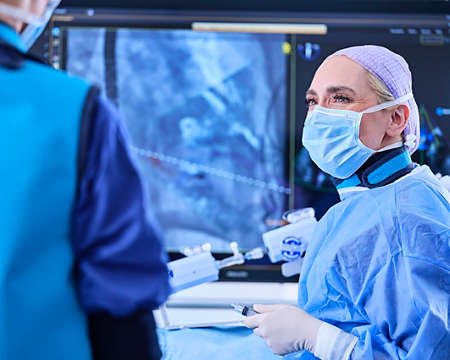
Preparation is everything when it comes to changing the landscape of minimally invasive structural cardiac interventions. The demands of transcatheter valvular and other structural heart procedures require a future-proof, fully integrated environment. Our SHD portfolio enables heart teams to grow their program and drive upcoming transcatheter innovations through workflow flexibility and multimodal imaging integration. This enhances communication, enables efficiency and provides confidence in anatomical guidance, device navigation, and accurate device placement.

Improving cath lab performance while staying close to the heart