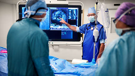
- Clarity and confidence
-
One system, many choices
Core supports a full suite of imaging and physiology analysis tools including FFR lesion assessment, iFR modality, iFR Scout, digital IVUS, high-resolution rotational IVUS, ChromaFlo stent apposition assessment, VH IVUS automatic tissue classification, and Pioneer Plus IVUS for peripheral procedures.* - iFR modality simplifies workflow
-
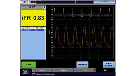
iFR modality simplifies workflow
iFR modality simplifies workflow by providing a hyperemia-free measurement to assess lesion significance in as few as five heartbeats. Philips’ proprietary iFR modality has a robust body of clinical evidence with over 9,000 patients in numerous studies and peer-reviewed journal articles.⁵ - iFR Scout pullback
-
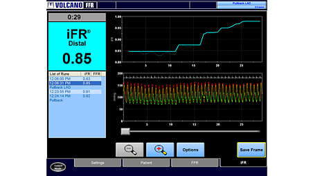
iFR Scout pullback
The iFR Scout pullback shows the most significant gradient in the mid-vessel lesion with diffuse proximal disease. - Fractional Flow Reserve
-
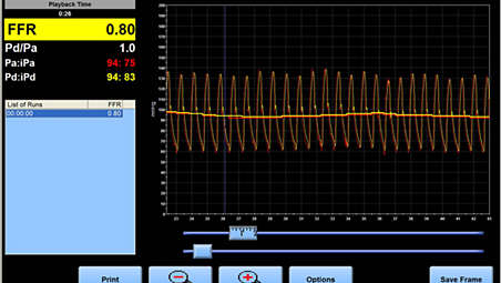
Fractional Flow Reserve measurement
Various clinical studies demonstrate that physiologic lesion assessment by FFR to guide routine PCI is superior to current angiography guided treatment. This measured ratio represents the potential decrease in coronary flow distal to the coronary stenosis.⁶ - IVUS assesses disease
-
IVUS helps with disease assessment
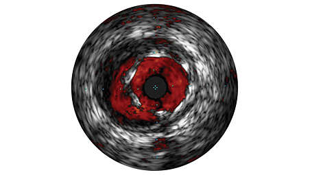
IVUS imaging helps physicians assess disease markers, including plaque burden percentage, lesion location and morphology, calcium volume, and the presence of thrombus. It also provides analysis of crucial parameters—like luminal cross-sectional measurements—and helps aid in disease diagnosis. - VH IVUS
-
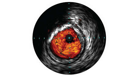
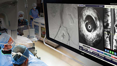
Real-time lesion assessment in the cath lab
VH IVUS Imaging provides a colorized tissue map of plaque composition with automated lumen and vessel measurements. VH IVUS technology uses advanced, proprietary spectral analysis techniques to classify plaque into 4 tissue types with 93-97% accuracy.⁹ - Grayscale
-
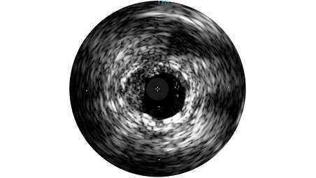
Grayscale enhances procedures
Grayscale enhances angiography procedures by enabling detailed views. Angiography produces a shadowgram of contrast, while IVUS visualizes extent and location of plaque, enabling precise disease assessment, vessel, and optimal stent placement. IVUS guidance has been associated with a 74% change in PCI strategy, and reduced MACE, MI, TLR, and death in large studies.⁷,⁸ - ChromaFlo
-
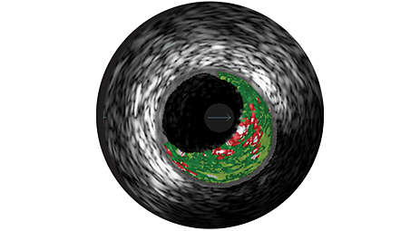
ChromaFlo stent apposition assessment
ChromaFlo highlights blood flow red for easy assessment of stent apposition, lumen size, and more. Appropriate for peripheral and coronary vessels, including left main, bifurcations, superficial femoral artery and iliac. It is designed to make lumen size and stent apposition instantly recognizable and helps identify branches, dissections, and plaque in bifurcations. - Convenience
-
True integration and convenience
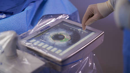
Always on and ready for use when you need it, Core delivers convenience in your workflow with true integration. Only Philips offers the plug-and-play simplicity of digital IVUS, touchscreen control from the sterile field to get to your answers faster, and the hyperemia-free iFR modality.²,³ - Optimal ease of use
-
Intuitive interface
Core delivers an intuitive interface for optimal ease of use as well as guided workflows and uniform controls to simplify training. Convenient measurements and labeling tools are available to document your findings. Core also offers a streamlined workflow with DICOM Worklist for patient data transfer. You can archive your results via DICOM store, DVD, or printout.²,⁴
One system, many choices
One system, many choices
One system, many choices
iFR modality simplifies workflow
iFR modality simplifies workflow
iFR modality simplifies workflow
iFR Scout pullback
iFR Scout pullback
iFR Scout pullback
Fractional Flow Reserve measurement
Fractional Flow Reserve measurement
Fractional Flow Reserve measurement
IVUS helps with disease assessment
IVUS helps with disease assessment
IVUS helps with disease assessment
Real-time lesion assessment in the cath lab
Real-time lesion assessment in the cath lab
Real-time lesion assessment in the cath lab
Grayscale enhances procedures
Grayscale enhances procedures
Grayscale enhances procedures
ChromaFlo stent apposition assessment
ChromaFlo stent apposition assessment
ChromaFlo stent apposition assessment
True integration and convenience
True integration and convenience
True integration and convenience
Intuitive interface
Intuitive interface
Intuitive interface
- Clarity and confidence
- iFR modality simplifies workflow
- iFR Scout pullback
- Fractional Flow Reserve
- Clarity and confidence
-
One system, many choices
Core supports a full suite of imaging and physiology analysis tools including FFR lesion assessment, iFR modality, iFR Scout, digital IVUS, high-resolution rotational IVUS, ChromaFlo stent apposition assessment, VH IVUS automatic tissue classification, and Pioneer Plus IVUS for peripheral procedures.* - iFR modality simplifies workflow
-
iFR modality simplifies workflow
iFR modality simplifies workflow by providing a hyperemia-free measurement to assess lesion significance in as few as five heartbeats. Philips’ proprietary iFR modality has a robust body of clinical evidence with over 9,000 patients in numerous studies and peer-reviewed journal articles.⁵ - iFR Scout pullback
-
iFR Scout pullback
The iFR Scout pullback shows the most significant gradient in the mid-vessel lesion with diffuse proximal disease. - Fractional Flow Reserve
-
Fractional Flow Reserve measurement
Various clinical studies demonstrate that physiologic lesion assessment by FFR to guide routine PCI is superior to current angiography guided treatment. This measured ratio represents the potential decrease in coronary flow distal to the coronary stenosis.⁶ - IVUS assesses disease
-
IVUS helps with disease assessment
IVUS imaging helps physicians assess disease markers, including plaque burden percentage, lesion location and morphology, calcium volume, and the presence of thrombus. It also provides analysis of crucial parameters—like luminal cross-sectional measurements—and helps aid in disease diagnosis. - VH IVUS
-
Real-time lesion assessment in the cath lab
VH IVUS Imaging provides a colorized tissue map of plaque composition with automated lumen and vessel measurements. VH IVUS technology uses advanced, proprietary spectral analysis techniques to classify plaque into 4 tissue types with 93-97% accuracy.⁹ - Grayscale
-
Grayscale enhances procedures
Grayscale enhances angiography procedures by enabling detailed views. Angiography produces a shadowgram of contrast, while IVUS visualizes extent and location of plaque, enabling precise disease assessment, vessel, and optimal stent placement. IVUS guidance has been associated with a 74% change in PCI strategy, and reduced MACE, MI, TLR, and death in large studies.⁷,⁸ - ChromaFlo
-
ChromaFlo stent apposition assessment
ChromaFlo highlights blood flow red for easy assessment of stent apposition, lumen size, and more. Appropriate for peripheral and coronary vessels, including left main, bifurcations, superficial femoral artery and iliac. It is designed to make lumen size and stent apposition instantly recognizable and helps identify branches, dissections, and plaque in bifurcations. - Convenience
-
True integration and convenience
Always on and ready for use when you need it, Core delivers convenience in your workflow with true integration. Only Philips offers the plug-and-play simplicity of digital IVUS, touchscreen control from the sterile field to get to your answers faster, and the hyperemia-free iFR modality.²,³ - Optimal ease of use
-
Intuitive interface
Core delivers an intuitive interface for optimal ease of use as well as guided workflows and uniform controls to simplify training. Convenient measurements and labeling tools are available to document your findings. Core also offers a streamlined workflow with DICOM Worklist for patient data transfer. You can archive your results via DICOM store, DVD, or printout.²,⁴
One system, many choices
One system, many choices
One system, many choices
iFR modality simplifies workflow
iFR modality simplifies workflow
iFR modality simplifies workflow
iFR Scout pullback
iFR Scout pullback
iFR Scout pullback
Fractional Flow Reserve measurement
Fractional Flow Reserve measurement
Fractional Flow Reserve measurement
IVUS helps with disease assessment
IVUS helps with disease assessment
IVUS helps with disease assessment
Real-time lesion assessment in the cath lab
Real-time lesion assessment in the cath lab
Real-time lesion assessment in the cath lab
Grayscale enhances procedures
Grayscale enhances procedures
Grayscale enhances procedures
ChromaFlo stent apposition assessment
ChromaFlo stent apposition assessment
ChromaFlo stent apposition assessment
True integration and convenience
True integration and convenience
True integration and convenience
Intuitive interface
Intuitive interface
Intuitive interface
Specifications
- Power requirements
-
Power requirements System input - 100, 120V, 220, 240VAC, 50/60Hz, 1000VA
Monitor - 100V – 240V 50/60Hz, 39W
Workstation - 100 – 240V, 50/60Hz, 550VA, 825VA
-
- Dimensions
-
Dimensions Control room controller - H=5'', W=15'', D=10'' Inches
-
- Ordering Information
-
Ordering Information Core system - CORE01
Control pad - CPADO1
Bedrail mount - MNTO1
Monitor mount - MNTO2
-
- Dimensions
-
Dimensions Workstation - H=17'', W=10'', D=16.5'' Inches
Control pad (optional) - H=2.75'', W=10.5'', D=8.3'' Inches
Monitor - H=15''– 19'' (depending upon the stand extension), W=15.8'', D=9.7'' Inches
Connection box - H=9.85'', W=2.95'', D=7.75'' Inches
-
- Power requirements
-
Power requirements System input - 100, 120V, 220, 240VAC, 50/60Hz, 1000VA
Monitor - 100V – 240V 50/60Hz, 39W
-
- Dimensions
-
Dimensions Control room controller - H=5'', W=15'', D=10'' Inches
-
- Power requirements
-
Power requirements System input - 100, 120V, 220, 240VAC, 50/60Hz, 1000VA
Monitor - 100V – 240V 50/60Hz, 39W
Workstation - 100 – 240V, 50/60Hz, 550VA, 825VA
-
- Dimensions
-
Dimensions Control room controller - H=5'', W=15'', D=10'' Inches
-
- Ordering Information
-
Ordering Information Core system - CORE01
Control pad - CPADO1
Bedrail mount - MNTO1
Monitor mount - MNTO2
-
- Dimensions
-
Dimensions Workstation - H=17'', W=10'', D=16.5'' Inches
Control pad (optional) - H=2.75'', W=10.5'', D=8.3'' Inches
Monitor - H=15''– 19'' (depending upon the stand extension), W=15.8'', D=9.7'' Inches
Connection box - H=9.85'', W=2.95'', D=7.75'' Inches
-
Related products
Alternative products
-
SyncVision
- iFR Co-registration
- IVUS Co-registration
- iFR and IVUS Tri-Registration
- Calibrated QCA
- Vessel enhancement
View product
-
Eagle Eye Platinum
- #1 choice of physicians for intravascular imaging (in the US).*
- GlyDx hydrophilic coating for increased lubricity
- A long, rapid exchange lumen for improved pushability
- Three radiopaque markers
- Compatibility with SyncVision for co-registration with angiography
View product
-
Eagle Eye Platinum ST
- A 2.5 mm tip-to-imaging distance designed to assess more of the vessel than standard catheters
- Plug-and-play simplicity
- Three radiopaque markers
- GlyDx hydrophilic coating
- SyncVision compatibility
View product
-
Verrata Plus
- New in-line clip connector for improved work flow
- New high fidelity sensor for reliable pressure measurement
- Reliably connect and disconnect with confidence
- Seamlessly switch between FFR and iFR, the only resting index with over 4500 patients studied
View product
-
Core Mobile
- Choice of FFR, iFR, and IVUS modalities on a single mobile platform
- Delivers convenience by offering flexibility to service multiple rooms
- Offers an intuitive interface for optimal ease of use
View product


-
SyncVision
The SyncVision precision guidance system is suitable with IntraSight and Core Integrated interventional platforms and streamlines lesion assessment, simplifies vessel sizing and enables precise therapy delivery all while integrating seamlessly in daily workflows in interventional suites of choice.
View product
-
Eagle Eye Platinum
The Eagle Eye Platinum digital IVUS catheter is the #1 choice of physicians for intravascular imaging (in the US).* As a unique plug-and-play intravascular imaging catheter it is designed for ease of use and deliverability. Features include a soft tapered tip, GlyDx hydrophilic coating for increased lubricity, a long, rapid exchange lumen for improved pushability, three radiopaque markers, and compatibility with SyncVision for co-registration with angiography.
View product
-
Eagle Eye Platinum ST
The Eagle Eye Platinum ST digital IVUS catheter offers a 2.5 mm tip-to-imaging distance designed to assess more of the vessel than standard catheters by providing a closer visualization of highly stenosed lesions and distal anatomy. The short tip catheter fits through all 5F guides and has all features of our top-selling Eagle Eye Platinum model, including plug-and-play simplicity, three radiopaque markers, GlyDx hydrophilic coating, and SyncVision compatibility.*
View product
See all related products -
Verrata Plus
For over 20 years Philips has led the way in physiology, putting patients first through continuous innovation. Now introducing our 10ᵗʰ generation physiology wire: Verrata Plus.
View product
-
Core Mobile
Core Mobile is designed to power the future of precision guided therapy and offers the choice of imaging and physiology on a single mobile platform.¹ Core Mobile also provides clarity in your approach and confidence in your results by offering additional information during the diagnostic phase, assisting in intervention decisions.
View product
- 1. 505-0100.21, Operator's Manual, Core Integrated, 3.4X (p. 18); 505-0101.16, Operator's Manual, s5 Series FFR_iFR Option, v3.4x (p.15).
- 2. VAL RPT, S5-Core V3.4 SW with Core Control Pad, 215-0007.02.
- 3. Product Spec. 809480-001, 202-0407.01.
- 4. Requirements Specification Meridian VH SW, 806000-004 (pg 84).
- 5. An iFR cut-point of 0.89 matches best with an FFR ischemic cut-point of 0.80 with a specificity of 87.8% and sensitivity of 73.0%. (iFR Operator’s Manual 505-0101.23) *Product availability is subject to country regulatory clearance. Please contact your local sales representative to check the availability in your country.
- 6. N Engl J Med. 2012;367(11):991-1001.
- 7. Witzenbichler B et al. Relationship Between Intravascular Ultrasound Guidance and Clinical Outcomes After Drug-Eluting Stents: The ADAPT-DES Study. Circulation 2014 Jan: 129,4;463-470
- 8. Ahn et al. Meta-Analysis of Outcomes After Intravascular Ultrasound Guided Versus Angiography-Guided Drug-Eluting Stent Implantation in 26,503 Patients Enrolled in Three Randomized Trials and 14 Observational Studies. Am J Cardiol 2014; 113:1338-1347